ED-7-4
Neutron transmission imaging system with a superconducting kinetic inductance detector
10:00-10:15 30/11/2023
*The Dang Vu1,2, Hiroaki Shishido3, Kazuya Aizawa2, Takayuki Oku2, Kenichi Oikawa2, Masahide Harada2, Kenji M. Kojima4, Shigeyuki Miyajima5, Kazuhiko Soyama2, Tomio Koyama1, Mutsuo Hidaka6, Soh Y. Suzuki7, Manobu M. Tanaka8, Masahiko Machida9,Shuichi Kawamata1 and Takekazu Ishida1
1. Division of Quantum and Radiation Engineering, Osaka Metropolitan University, Sakai, Osaka 599-8570, Japan
2. Materials and Life Science Division, J-PARC Center, Japan Atomic Energy Agency, Tokai, Ibaraki 319-1195, Japan
3. Department of Physics and Electronics, Graduate School of Engineering, Osaka Metropolitan University, Sakai, Osaka 599-8531, Japan
4. Centre for Molecular and Materials Science, TRIUMF, 4004 Wesbrook Mall, Vancouver, BC V6T 2A3, Canada
5. Advanced ICT Research Institute, National Institute of Information and Communications Technology, 588-2 Iwaoka, Nishi-ku, Kobe, Hyogo 651-2492, Japan
6. Advanced Industrial Science and Technology, Tsukuba, Ibaraki 305-8568, Japan
7. Computing Research Center, Applied Research Laboratory, High Energy Accelerator Research Organization (KEK), Tsukuba, Ibaraki 305-0801, Japan.
8. Institute of Particle and Nuclear Studies, High Energy Accelerator Research Organization (KEK), Tsukuba, Ibaraki 305-0801, Japan
9. Center for Computational Science & e-Systems, Japan Atomic Energy Agency, 178-4-4 Wakashiba, Kashiwa, Chiba 277-0871, Japan.
The discovery of X-Ray [1] laid the foundation of the non-destructive and non-invasive technique, which has a wide range of applications in medical and material sciences. However, we consider that neutron beams have a superiority over X-Rays because they are able to deeply penetrate into electron cloud not only to interact with nuclei as particles [2] but also to probe spin-magnetic fields in materials. Therefore, in combination with X-ray imaging [3], the neutron imaging technique has been prospected as an indispensable tool in the field of material sciences [4] for reinforcing the usefulness of the non-destructive and non-invasive technique [3,4].Currently, the best spatial resolution of the neutron imaging was reported as 2 µm using scintillation neutron detectors, while a detection efficiency remains to be lower than 1% [5]. Alternatively, a semiconductor neutron detector or a solid-state neutron detector has an advantage in its small size and a low operating voltage, and is profitable to use for the use of time-of-flight measurements, but it is not the best when considering the its spatial resolution of 15 µm and its detection efficiency low at 2% [5]. One of the outstanding detectors is supposed to be a 10B-doped microchannel plate (MCP). It has a high spatial resolution 15 μm at low count rates and 55 μm at high count rates, and a high detection efficiency of 43% for cold neutrons and 16% for thermal neutrons [5].
Our group proposed a current-biased kinetic inductance detector (CB-KID) system as a new superconducting neutron detector [6]. The CB-KID is different from others because it is based on a rapid reduction of the Cooper pair density nsat a local tiny spot of the nanowire stripline in a micrometer length Δℓ (≪ℓ) when a neutron reacts with a 10B nucleus in the conversion layer. Under feeding modest DC bias currents, two pairs of voltage pulses appear at the holt spot and propagate along two superconducting striplines which are fabricated orthogonally with each other.
The delay-line technique is a conventional method to obtain a neutron transmission image in two dimensions using delay-line timestamps from the four terminals of CB-KID. In contrast with the independent-pixel-based imaging in our earlier approaches in combination of CB-KID and the readout circuit composed of superconducting digital devices [6], a superconducting delay-line method was first planned in a proposal (JP16H02450) by the leader in 2015 by utilizing a time-to-digital converter of the readout circuit named as Kalliope-DC [7]. Based on systematic studies on the characteristics of the CB-KID detector [8,9]. Meantime, the operating conditions have been optimized for improving the spatial resolution, the temporal resolution, and the detection efficiency. In addition, various different samples have been examined to demonstrate the usefulness of our CB-KID system in wide-field applications, i.e., a good linearity between the Gd-island sizes in neutron imaging and SEM images over the wide range of sample sizes [10], the possibility in obtaining the narrow-area Bragg-edge neutron transmission [11], the Bragg-dip imaging of small YbSn3 single crystallites [12], and the confirmation of spatial resolution down to 16 μm [13]. We recently built a superconducting neutron cryostat with a concaved flange in the upstream side of the CB-KID detector to mount/replace samples at room temperature at a distance of 20 mm to sensor [14]. This enables the following things to be done, i.e., an easy change of the sample environments, the measurements of larger samples, and a space available to install the neutron tomography system. An open-beam normalization became possible during the same beam time because of its importance in correctly obtaining neutron-transmission spectra. For example, a sequential change in microstructure of Wood’s metal alloy [14] was observed successfully by melting and re-solidification of the alloy during the same beam time at J-PARC (MLF). We also observed not only Bragg-dips in neutron transmission spectroscopy from CaF2, FeS2 but also neutron resonance dips from Ti and V during the same beam time.
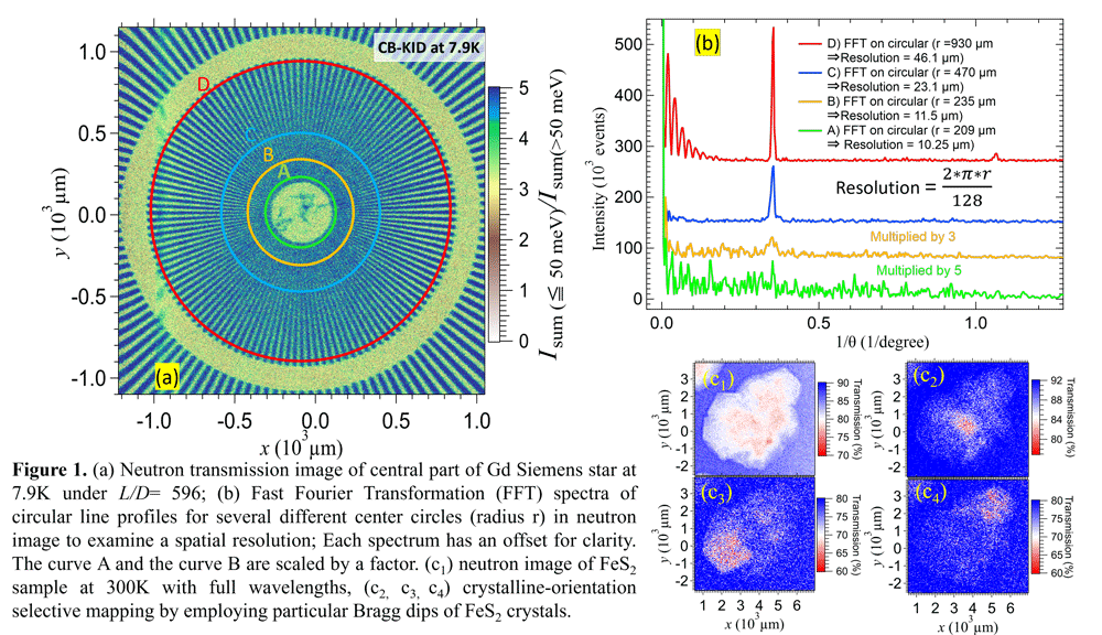
In this study, we evaluated a change in spatial resolution of neutron-transmission image between the cryogenic-environment mounting and the room-temperature mounting of a sample. We have to choose the operating conditions of neutron detector and a neutron-collimator setting to satisfy an experimental purpose. For example, we have to consider whether the best-spatial resolution is needed or the data statistic is more important in each case. Note that our system is able to mount a sample either at a cryogenic temperature or at a room temperature for neutron transmission imaging. We took different radius circular line profiles of Gd Siemens-star (128 spokes) pattern (Fig. 1(a)) in neutron-transmission image and applied the fast Fourier transformation (FFT) to check the appearance of fundamental peak. We consider that a minimum discernable period of the Gd spokes is a good measure of resolution. Operating condition and collimator ratio (L/D) of beams were appropriately chosen so as to meet the purpose. We found a spatial resolution ~11 µm when the Gd Siemens star was placed at a cryogenic temperature under a rotary collimator “Small” (L/D = 596) (see Fig. 1(a), (b)). We also confirmed that our CB-KID sensor is able to observe the neutron transmission coefficient over a wide range of neutron energies. Using a natural FeS2 single crystalline sample placed at room temperature, we observed the Bragg dips caused by FeS2 crystals in the transmission spectrum. We succeeded in mapping differently-oriented crystals by choosing several particular Bragg dips of FeS2 crystals to compose images (seeFig. 1 (c1, c2, c3, c4) and in assigning nuclear resonance dips with those predicted from database cross-sections up to higher energies to 100 keV.
[1] Röntgen W C Nature 53 (1896) 274–6
[2] Tengattini A, et al., Geomechanics for Energy and the Environment 27 (2021)100206–19
[3] Tengattini A, et al., Nucl Instrum Methods Phys Res A 968 (2020) 163939
[4] Kardjilov N, et al., Materials Today 21 (2018) 652–72
[5] Pietropaolo A, et al., Phys Rep 875 (2020) 1–65
[6] Ishida T, et al., J Low Temp Phys 176 (2014) 216–21
[7] Kojima, K. M., et al., J Phys Conf Ser 551 (2014) 012063.
[8] Vu T D, et al., J Phys Conf Ser 1293 (2019) 012051
[9] Vu T D, et al., J Phys Conf Ser 1590 (2020) 012036
[10] Vu T D, et al., Nucl Instrum Methods Phys Res A 1006 (2021) 165411
[11] Vu T D, et al., J Phys Conf Ser 2323 (2022) 012028
[12] Shishido H, et al., J Appl Crystallogr 56 (2023) 1-6
[13] Iizawa Y, et al., Supercond Sci Technol 32 (2019) 125009
[14] Vu T D. et al., J Phys Conf Ser 2545 (2023) 012019
This work is partially supported by Grants-in-Aid for Scientific Research (JP16H02450, JP21H04666, JP21K14566, and JP23K13690) from JSPS. The neutron irradiation experiments at MLF of J-PARC were conducted under the support of the MLF project program.